
There are many variations on pileipellis structure, which are here simplified into four main types.
The universal veil may consist of quite different elements to the underlying pileipellis, such as in some species of Amanita, where the veil is largely of globose elements overlying a cutis. If there is a clearly distinct universal veil which leaves remnants on the pileus surface, then determine the pileipellis structure from the underlying pileus surface. However, in some cases it is difficult to distinguish the hyphae of the universal veil from those of the pileus surface. In such cases we treat the whole outer layer as the pileipellis, irrespective of the origin of the hyphae.
Differentiated Pileocystidia may project from the pileipellis.
Terminal elements and the hyphae of the pileipellis may be branched in various ways: see Pileipellis terminal elements (surface) and Pileipellis hyphae (branching).
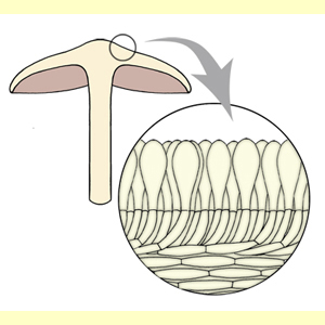
A tangential cross section will be similar to the radial cross section, as far as the orientation of the surface elements. A scalp section will show elements with a globose to subglobose outline (looking down on the projecting ends of the hyphae).
Where the terminal elements arise at different levels, the structure is a trichoderm. Transitions to an epithelium are possible, with inflated elements mostly arising at the same level, but some not. Choose both hymeniderm and epithelium for such a structure.