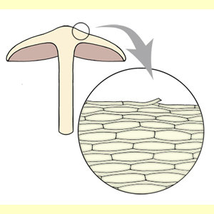
The pileipellis is the outer layer of the pileus, above the trama (flesh). The structure of the pileipellis is determined from a radial cross section about half-way between the pileus centre and edge. A scalp section from the same area may be useful in interpreting the type and arrangement of the elements that make up the pileipellis. The pileipellis may be uniform or layered. If layered, structure refers to the outer layer. The lower layer, next to the trama, is the hypoderm, which may have hyphae of different shape or gelatinisation to the upper layer.
There are many variations on pileipellis structure, which are here simplified into four main types.
The universal veil may consist of quite different elements to the underlying pileipellis, such as in some species of Amanita, where the veil is largely of globose elements overlying a cutis. If there is a clearly distinct universal veil which leaves remnants on the pileus surface, then determine the pileipellis structure from the underlying pileus surface. However, in some cases it is difficult to distinguish the hyphae of the universal veil from those of the pileus surface. In such cases we treat the whole outer layer as the pileipellis, irrespective of the origin of the hyphae.
Differentiated Pileocystidia may project from the pileipellis.
Terminal elements and the hyphae of the pileipellis may be branched in various ways: see Pileipellis terminal elements (surface) and Pileipellis hyphae (branching).
Choose this state if: in radial cross section the outer layer of the pileipellis consists of repent (parallel to the surface), cylindrical or fusiform hyphae. Hyphae are typically arranged radially and more or less parallel to one another (parallelocutis), but are occasionally not arranged radially or parallel to one another (mixtocutis,
Dryophila type cutis). Some hyphal ends may curl upwards from the surface, but the terminal elements are not all oriented vertically (as in a
trichoderm). This type of pileipellis is often associated with a silky or radially fibrillose macroscopic appearance of the pileus.
Hyphae may occur in a gelatinous matrix (an ixocutis).
A tangential cross section will reveal the more or less circular cut ends of hyphae. A scalp section will show radially arranged hyphae.

